The genus Lacrymaria contains species with dark brown gills that are very similar in colour to Agaricus. Today I spotted the mushrooms below on the side of the road in the middle of Karri forest near Pemberton in SW WA.
Lacrymaria velutina
The gills were a dark chocolate brown.
Gills of Lacrymaria velutina
From the above shot a couple of things stand out. Firstly, the gills are attached to the stipe. This differs from Agaricus where the gills are always free. Secondly, the stipe has split into a number of strips. This does not happen with Agaricus.
A close-up of the gills also shows that they are blotchy in appearance, which we do not seen in Agaricus.
Close-up of Lacrymaria velutina gills
Under UV light the entire mushroom glows a lovely purple colour. Unfortunately I was unable to capture this colour on my iPhone.
Note: I have named this specimen from online images. It may be a native species that is not described.
Lacrymaria are not considered to be edible mainly because of taste rather than toxicity. It has a very thin flesh in any case so would not make much or a meal.
Within the genus Agaricus, the Section Xathodermatei contains a number of species that are commonly known as yellow stainers and they are known to contain phenol which causes quite nasty gastric upsets if consumed. I have been meaning to put together a post about these but it was only this morning that I found a substantial patch of them on a street verge to do some images and experiments. A few members of the patch are shown in the picture below.
Yellow stainers on street verge
The yellow staining reaction is seen both on the cap and on the stem of the mushrooms and manifests itself as a bright chrome yellow stain that quickly fades. Once picked, the yellow stain on the cap may not continue to show itself. The picture below shows the sort of stain that occurs when you first pick one of these mushrooms. This was completely gone within 2 minutes.
Yellow stain on edge of cap when first picked
The partial veil on these mushrooms has a fluffy appearance that I believe can be called flocculose. The appearance of the partial veil at various stages is shown in the following set of images. The centre image shows both the partial veil and the remains of the universal veil. Together, these form the ‘double annulus’ that is a characteristic of this Section.
Partial veil at various stages of growth
The yellow stain on the cut stem can also been seen in these images as can the white core in the centre of the stem, a feature that is also seen in supermarket mushrooms.
Another feature that tends to be a characteristic of mushrooms in this Section is the ‘boxy’ cap shape. That shape can be seen in the first image above. This is where the analysis gets interesting. As I have mentioned elsewhere, the yellow stain can be made permanent on these mushrooms by applying an alkali. The yellow colour is due to 4,4-dihydroxyazobenzene. While looking closely at these mushrooms I noticed that they have a very distinct internal structure featuring a very dense section in the cap above the stem. This is revealed in a sectioned piece developed with Napisan solution which provided the necessary alkalinity and perhaps some oxidizing power that might have had an effect. An example of a sectioned mushroom developed in this way is shown in the pictures below, compared with a supermarket version.
Yellow stainer (top) compared with normal supermarket mushroom (bottom), both developed with Napisan solution
In this view it is immediately apparent that there is a significant difference in the internal structure of the two mushrooms. The hard core in the centre of the cap of the yellow stainer shows up clearly. I strongly suspect that this structure is responsible for the boxy shape of these mushrooms. Beyond that however, the flesh of the cap of the yellow stainer remains refractory to the effects of the Napisan while the flesh of the supermarket mushroom saturates and slightly darkens. The hard core also influences the way the cap separates from the stem. In the case of the supermarket mushroom the separation is very clean but in the case of the yellow stainer the stem breaks away with a rather ragged edge, as shown in the picture below with two yellow stainers on the right and two supermarket varieties on the left.
Cap separation on supermarket mushrooms (left) versus yellow stainers (right).
I have in mind a few more experiments on these interesting mushrooms but I will finish this post off for now.
A quick postscript. After about an hour the difference between the two mushrooms became even more stark.
I found this monster growing at the base of a eucalyptus tree in Bridgetown. It was almost buried, rather like Agaricus bitorquis. Although I have called it Agaricus osecanus, that is a fairly loose term, referring to a group of similar mushrooms. I am using Arora as a guide.
I first found these during Spring, and then again in Autumn. The most distinctive feature of them is the huge diameter of the stem. As you can see, it is a handful. In fact the specimen above appears to be three individuals fused together and it had dried out a little and cracked so that it was in danger of falling apart. The flesh however was quite firm.
I was quite excited when I found such a large mushroom that was clearly an Agaricus of some sort and therefore likely to be edible. I thought that I would take a culture of it as soon as a I could. However, the smell was not like any other member of the genus that I have ever encountered. A friend described it as ‘earthy’ but I found it simply ‘less than attractive’.
Before going to the trouble of culturing it, I decided to do a small taste test. To this end, I cut some small slices (no colour change) and fried them in a little butter/oil. My friend and I both tasted it and though the initial taste seemed ok, we had both spat it out within 5 seconds. It tasted terrible. Hard to describe exactly, but I found it to have a floury taste. Certainly not something that you would want to swallow.
So, for me this represents a fourth grouping within Agaricus, based mainly on the smell. The other three categories are mushroom (octenol) smell, almond smell and phenol smell. I can’t put a description to this smell, but it does not fit into any of the other three categories. `
Such a shame. It was massive and had lovely white firm flesh.
Ah well.
Note: May 2016
I had hoped to get another specimen of this and send it off for dna testing, but this year someone came along and not only smashed them up, but ripped off all the loose bark of the tree whose base it was growing at. Such a senseless act and now we may never know what this was.
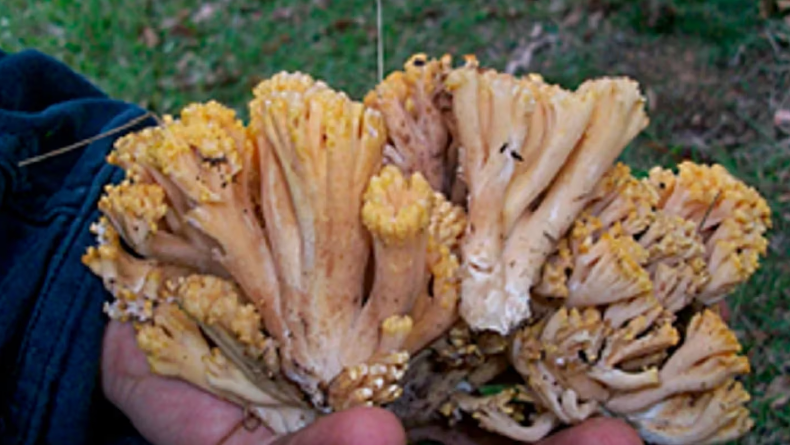
I first became interested in Ramaria ochraceosalmonicolor after the eminent Naturalist J. H. Willis mentioned that he had eaten it in his 1957 publication ‘Victorian Toadstools and Mushrooms’. Ramaria are not easy to identify and any perusal of the internet will find various illustrations with this name but looking nothing like the picture below. There is even a paper in the Australian Journal of Mycology (2007) which goes into much detail about the naming of the species.
For my purposes, however, the important thing was to establish what Willis had eaten. The paper linked above mentions that Willis as well as Bougher and Syme show illustrations of a coralloid structure for this fungus. Though the Bougher and Syme illustration is clear and matches the photograph, I was puzzled by the reference to Willis until I noticed that he had an illustration of three species of Ramaria as a fronticepiece in his book. I had previously overlooked these illustrations. Comparison with his images left me in little doubt that this is the form of Ramaria that he was referring to.
This being the case, I set some aside for a sampling. Ramaria can be risky, with a tendency to cause diahorrea according to Arora, so I decided to set them aside in the fridge and try them in the morning, rather than risk and uncomfortable night.
To be continued….
Ramaria ochraceosalmonicolor
So, I fried up the sample that I had collected and consumed about 2 tablespoons full at 10:30 in the morning. It is now 6:45 in the evening and I have had no reaction. But what an anxious time it has been. After consuming the fungus, I began googling and came up with Ramaria flavo-brunnescens. It grows exclusively under Eucalyptus in Brazil and other places in South America, and has been responsible for the death of cattle. There is a report with gruesome histological details. I am at a loss however to understand why there are not similar reports from Australia, given that there must be many cattle grazed where there are Eucalypts.
In Uruguay it grows in profusion in April, May and June where it causes a disease known as bocopa in sheep, cattle, pigs and horses. The disease has high mortality rates and is increasing in extent.
Ramaria flavo-brunnescens
There is, futhermore, a report of human poisoning and death from this fungus (the same one as in Brazil), although admittedly in combination with an Amanita, from China.
The images from the Brazilian report are disturbingly similar to my image above. Certainly enough to be within the general area, and the reference to Eucalyptus is especially unsettling. The poison is unidentified. It is reported to affect the incorporation of sulphur-containing amino acids such as cysteine. It is also most likely volatile, as toxicity is not present in dried samples.
The books in my library vary in their assessment of this fungus. Willis says he has eaten it. Kevn Griffiths says it upsets some people, Bougher and Syme declare it poisonous.
In the balance, I suggest that this fungus is far too difficult to identify to consider it edible and there is some potential for it to be lethal. Despite my experience of consuming a small portion of a cooked specimen and surviving, I suggest that it be considered an inedible species.
While there is a tantalising morsel about the toxin in google books, the key information is an orphan on an invisible page and I am loathe to spend the $137 necessary to purchase the entire book online. It simply is not worth the bother. The reward is not worth the cost. I post this report so that there is at least some documentation on the internet regarding this genera. I cannot find a single report of the progress of poisoning by this genus in humans on its own available on the internet. Neither do my books on poisonous fungi describe the progression of the syndrome.
30 April 2015
There are more Ramaria out at the moment with a wide variety of colours and forms. It prompted me to have another look for references and I found this one with some images that clearly show the ‘cauliflower’ form that is supposed to be a characteristic of this species. Note however the slight difference in nomenclature.
I haven’t seen any specimens this compact, but I have consumed one of the local species with a local man of Italian descent. I have made a video of him with it that I will process and upload when I have time.
If anyone has a link or relevant experience, I would appreciate hearing about it.
A species which is frequently encountered, particularly around chicken coups is Chlorophyllum brunneum, pictured above. (You may see this same picture in a book, used without permission and mis-labelled) This is distinguished by the basal bulb which is described as ‘abrupt’, which means that there is usually a definite change in geometry at the top of the bulb and it often has quite a flat top. They often cluster together from a common large basal bulb as shown below.
It also has a surface that is broken up into scales that have a fibrous appearance. When cut it turns red.
Personally I find Chlorophyllum brunneum to be a very tasty mushroom with a strong meaty smell and flavour. But apparently it does not agree with some people so caution is wise. There is some reason to believe that bad reactions can be avoided by making sure that the mushroom is well-cooked. That said, I am aware of one report where someone who has eaten these on multiple occasions suddenly suffered some very unpleasant reactions after preparing it in their customary manner. It is possible that this mushroom contains a toxin similar to Chlorophyllum molybdites but of lower amount or strength. Since this is a protein toxin with a known molecular weight it should be possible to determine this by electorphoresis.
Although this mushroom is similar to Chlorophyllum rachodes, we are advised by mycologist Else Vellinga that that species does not occur in Australia. Here is a link to Vellinga’s paper. It does, however, occur in New Zealand and has been identified there by Mycologist Jerry Cooper. The two species can be difficult to tell apart without some careful microscopy.
Below are another couple of images of Chlorophyllum brunneum. The first shows a young fresh specimen and the other shows a close-up of the gap between the gills and the stem where a green ring can be seen.
This tale of poisoning by my friend Martin is reproduced with his permission
I post and record this experience here so others won’t make the fatal mistake I did but also to potentially kill off some of my mushroom ego if you like
So as some of you may know I escaped the mainland last week on the last flight into tassie to go bushwalking with my wife who is not my wife who I would like to be my wife and step daughter etc both to Bush walk but secretly to hunt down in my opinion what I have heard to be the worlds greatest tasting mushrooms – and later I will post other things about various finds and it was really tongue to ground amazing but the story I am about to share was not so amazing well actually it was kind of amazing – you can see from the above grammar is not my strong point
So I picked some C brunneum (pictured) before the 7 day hike – and they were growing on mass like most mushrooms in Tasmania do – and on day one of the hike I got lost and a 7 hour walking day turning into an 11 hour walking day so when I arrived to camp I got out a bottle of wine and the Brunneys – Now I have eaten these mushrooms on three previous occasions and they are marvellous – so I told the rest of the camp the stories of hunting them down and cooked up a batch on high heat in the pan and surprisingly everyone except one fellow took my word for it and ate them – I was surprised as most people I find not so open to new mushrooms – but my banter must have been right on point cause everyone agreed a great tasting mushroom – and at this point I was the mushroom king
But this is where the story takes a twist
My wife who is not my wife but I would like to be wife offered to cook the next batch as I was tired and dead from walking and getting lost and mentioned something like I will cook these with less oil and low heat to save on gas etc and by this time I was tipsy and ranting about black trumpets and finding the lost porcini of Tasmania etc that I didn’t give it a second thought and a very large plate of mushrooms shortly arrived in front of me warm and a little on the raw side which I devoured very quickly with waving arms and dancing.
Fast forward a few hours later in my sleeping bag and I turn to my wife who you know isn’t my wife and she has ear plugs in and i say I feel sick and she says you always say that after eating mushrooms etc.
I just make it out the door and I am projectile vomiting all over the forest and everybody can hear the mushroom king – and this vomiting goes on and off for the next hour and it’s minus 3 outside and my wife is yelling at me also and nothing is subtle here – maybe no one can hear me I think – after cleaning up and hiding all the spew and evidence I think maybe no one will catch on And I make it back to bed.
After an hour or so there are indescribable sounds coming from my belly – and I should probably wind it up here but I really want to crush a good part of my mushroom ego here so I will continue – I find myself in thermal onesie which happens to be inside out
And I am listening to the sound in my belly going wow that sounds very impressive and then I think I need to get out of here quick
I make it to a tree and can’t find the zip as it not on the outside and what unfolds now is many folds and it’s too late to stop it and it’s coming out my ankles
The rest of the details and screams and tears into the frozen night is not that important here the clean up in frozen river etc
Over the next 6 days I found amazing edible mushrooms but I was now on a ban – my wife who is not my wife who I want to marry who won’t currently marry me has banned me from eating mushrooms and I had lost my title of the mushroom king – but over the next days I would find fields of giant laccaria and 10s of thousands of golden chanterelles and wild enoki and fist sized hedgehogs and I was banned
And no one believed anything I said at the camp – I was reduced to nothing and a laughing stock – but secretly I was happy to have lived through it cause you need some setbacks to clarify where you are headed in life don’t you think ?
The ban was lifted when on the 7th day the chants appeared thank the lord – and I am alive to tell the tale – I remember shouting into the night my kidneys are going to explode get a helicopter here now to Rosie my wife who is not
So what to take from all this –
Peter Donecker will probably be able to tell me what chemical I ingested in the brunneum and the importance of making sure chlorophyllum is well and truly cooked at a high heat etc before sticking them down the throat
The good news is I am alive
And have been eating many mushrooms and have been taken down a fair few pegs
But haven’t eaten Brunneum again
But I will be in Melb on Tuesday so hoping to find some.
The spores of Chlorophyllum brunneum are approximately 10.1 x 6.5 microns. In the image below they are shown at 1000x magnification under oil with lactophenol blue stain.
Spores of Chlorophyllum brunneum.
Chlorophyllum molybdites
Another member of the genus, Chlorophyllum molybdites (below) has a very similar appearance, but the scales do not have the fibrous nature of C. brunneum. The gills start out white then gradually turn green. This green colour becomes much more pronounced as the specimens age and eventually the gills become very dark grey-green. It also has a green spore print when mature.
Chlorophyllum molybdites showing green gills
The spores have a green colour that can be seen from a spore print.
Green spore print of Chlorophyllum molybdites
It is sometimes claimed that this mushroom does not stain red, but this picture shows that it does indeed give a red stain when cut in two. Not quite as vivid as Chlorophyllum hortense but undoubtedly red.
Red colour of cut stem
The lower half of the stem on this mushroom has dark shading. This can vary a little in intensity. The annulus has two edges. These features are seen on the next image.
View showing gills and annulus
Sometimes the scales are almost absent as in this specimen from a verge in suburban Perth where it is sometimes particularly prevalent in periods of high temperatures and wet conditions.
It is not a deadly mushroom, but it may make you very sick and is a common cause of mushroom poisoning in North America. The nature of the poison in this mushroom was a mystery for a long time, particularly since it doesn’t affect all people at all times. It was revealed in 2012 to be a protein called molybdophyllysin by Yamada et al. It is heat labile, beginning to break down at 70 degrees, which may explain why some people, including the Cribbs report having eaten C. molybdites without ill effect.
The effect of temperature on the toxin in Chlorophyllum molybdites is shown in this graph from the above mentioned paper which plots activity against temperature for a 10 minute hold time. It might be anticipated that prolonged boiling might considerably reduce toxicity. There is similar information on the heat lability of this mushroom in a 1974 paper by Eilers and Nelson where it is referred to by an earlier name, Lepiota morganii. They report extraction of the toxin with different solvents, water being the most effective. One of these days I will take this data and try to concoct an equation for the reaction kinetics. They also mention that the toxin occurs in both young and mature specimens and in all parts of the fruiting body. This snippet of information eliminates a hypothesis that the toxicity is related to the stage of development of the mushroom.
Molybdophyllysin is a zinc metalloprotease. There are many of this type of enzyme in the biosphere, some toxic and some not. For example, Grifola frondosa, the famed ‘Maitake’ mushroom contains an enzyme of this class. It has found application as a tool for protein analysis as it cleaves proteins in a very specific way. On the other hand the toxin from Clostridium botulinum, the toxin that causes botulism is one of the most potent toxins known. Many studies have been done on the thermal stability of botulinum toxin and it is interesting that the recommended cooking time is a minimum of 5 minutes at 85 degrees centigrade. It would appear that the degradation of molybdophyllysin (MP) may follow very similar kinetics. One difference however is that MP is more susceptible to acidic conditions.
As a footnote, there is a report of the effects of eating this mushroom in the Medical Journal of Australia, by local academic Lindsay Mollison. I note that his report is in December 2011 and that he speaks of doing an extensive internet search to find out what he had eaten. Perhaps his experience was just prior to when I made this original post in July 2011. A shame. The first publication scientific publication describing this mushroom from Perth was written by mycologist Neale Bougher in
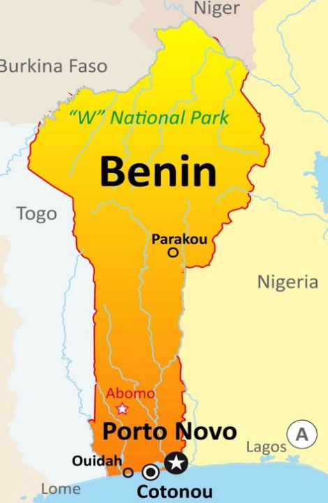
Although Chlorophyllum molybdites is considered poisonous in most places, there are reports of it being eaten in various places, particularly in Benin, in Africa. If you don’t know where that is, I have included a map below.
Here is a quote from a long treatise on the edible mushrooms of this country.
“In the area where we worked, there is no information on cases of poisoning caused by ukulé malu, nan bisu or bela-dedji. Benin is apparently only occupied by an edible form or with a low toxicity rate. It is also found that Chlorophyllum aff. molybdites is known and appreciated by the Peuhls, an important ethnic group that crosses the entire Sudano-Guinean region of West Africa. Finally, we point out that in Benin, mushrooms, with a few rare exceptions, are always eaten after preparation, that is to say after warm and relatively long heating. If the Chlorophyllum from Benin contained a labile toxin (thermolabile), it would be systematically destroyed by cooking or by blanching. It is clear that further toxicity and taxonomic studies will be needed to clarify and understand the toxicity of Chlorophyllum. For this reason we identify all our collections provisionally as Chlorophyllum aff. molybdites.
This small cluster of mushrooms appeared recently in a garden bed amongst some horse manure.
These mushrooms have a white cap with a brown colouration in the middle and when we flip them over, we can see that the gill colour is in the right range.
However, if we try to separate the cap from the stem, we find that we can’t, and the stem is furthermore completely hollow and thinner than what we might expect from an Agaricus. The mushroom pictured is Candolleomycescandolleana. It was formerly in the genus Psathyrella candolleana and some books list it as such. There is another species more commonly seen in forests called P. aspersopora. Both are of unknown or doubtful edibility though there is no record of them poisonous.
During another walk this evening I encountered another mushroom that had a similar appearance to an edible field mushroom. I picked it and brought it back to the house to document why it is not an edible field mushroom. Here is a picture of the cap.
Hebeloma westraliense, showing glutinous cap
The cap is not outside of the colour range that one might expect for an edible field mushroom, but notice that it is shiny? In fact it is quite slimy to the touch. This alone is enough to declare it to not be an edible Agaricus. However, let us continue…
Hebeloma westraliense, showing gills and top of stipe.
When we flip the mushroom over, we can see that the gills are in the right kind of colour range and that the stem has the right sort of thickness in relation to the cap. In fact, the gills even darken from pinkish to brown over time. However, the thing that is glaringly absent is an annulus or ring on the stem. Not a hint of one! We know for sure now that this is not an edible Agaricus, but lets go further…
Hebeloma westraliense, torn
If we attempt to snap the stem away from the cap, the result is unsuccessful. The whole cap tears apart rather than breaking at the junction of the stem and the cap. There is no change in the tissue type between the stem and the cap. This thing has now failed three tests. Quite a pretty mushroom never the less 🙂
If you are wondering about the tabletop, it is Australian red cedar, Toona ciliata. The mushroom is Hebeloma westraliense, edibility unknown (Bougher and Syme). Hebelomas are very useful for promoting the growth of Eucalypts and are cultivated for that purpose worldwide.
December 2023
In pine forests, and associated with introduced trees there is another species called Hebeloma crustuliniforme, also known as Poison Pie that is known to be poisonous.
Hebeloma crustluniforme with $10 note for size comparison.
Hebeloma crustuliniforme, torn in half.
I often hear people offer the advice “if you can peel a mushroom it is edible”. Of course such generalisations are nonsense, since you can peel a death cap. But I suspect that the origin of this advice is that this is a means of distinguishing between Agaricus and Hebeloma.
I was walking along the fire track this evening when I spotted this lovely big white mushroom cap, about 100 mm across.
Now, in colour and shape, this is not disimilar from an edible Agaricus. It is similar for example to Agaricus bitorquis. The location, in undisturbed eucalypt forest is a little unlikely for an Agaricus such as that however.
It can be seen that the edge of the cap has remnants of a torn veil. This might lead one to think that it is an Amanita. However, on flipping the mushroom over, all is revealed.
We can see from this that the mushroom has a ring on the stem, like an edible Agaricus and that the gills are within the right colour range, but on the surface of that ring is a deposit of orange spores. This shows us immediately that what we are looking at is not an Agaricus, but is in fact a Cortinarius of some kind. If it was an Agaricus, it would have a chocolate brown spore print.
Further differentiation from Agaricus is provided by the fact that the stem and the cap are part of the same structure. In Agaricus, the stem will break away cleanly from the cap.
As a rule, species in the genus Cortinarius are poisonous, so this is one mushroom that we would definitely avoid!